

The gap is real.
And it's preventable.
Female athletes tear their ACL 2 to 8 times more often than their male counterparts in the same sport. Understanding why is the first step to closing that gap.
Same sport. Very different risk.
In sports like soccer, basketball, and volleyball — where both sexes compete at high levels — female athletes consistently suffer ACL tears at dramatically higher rates. This isn't a coincidence or a matter of effort.
The disparity is rooted in biology, anatomy, and the way female bodies develop during adolescence. Hormonal shifts, skeletal geometry, and neuromuscular patterns all converge to create a window of elevated vulnerability — especially between ages 14 and 17.
The good news: most of these risk factors are modifiable. Targeted training can correct muscle imbalances, improve landing mechanics, and dramatically reduce injury rates — but only if athletes and coaches understand what they're up against.
ACL injury rate by sport
Relative injury rates per 1,000 athlete-exposures. Source: NCAA Injury Surveillance Program.
Built differently
Several anatomical differences in the female body create structural conditions that place greater stress on the ACL during everyday athletic movements.
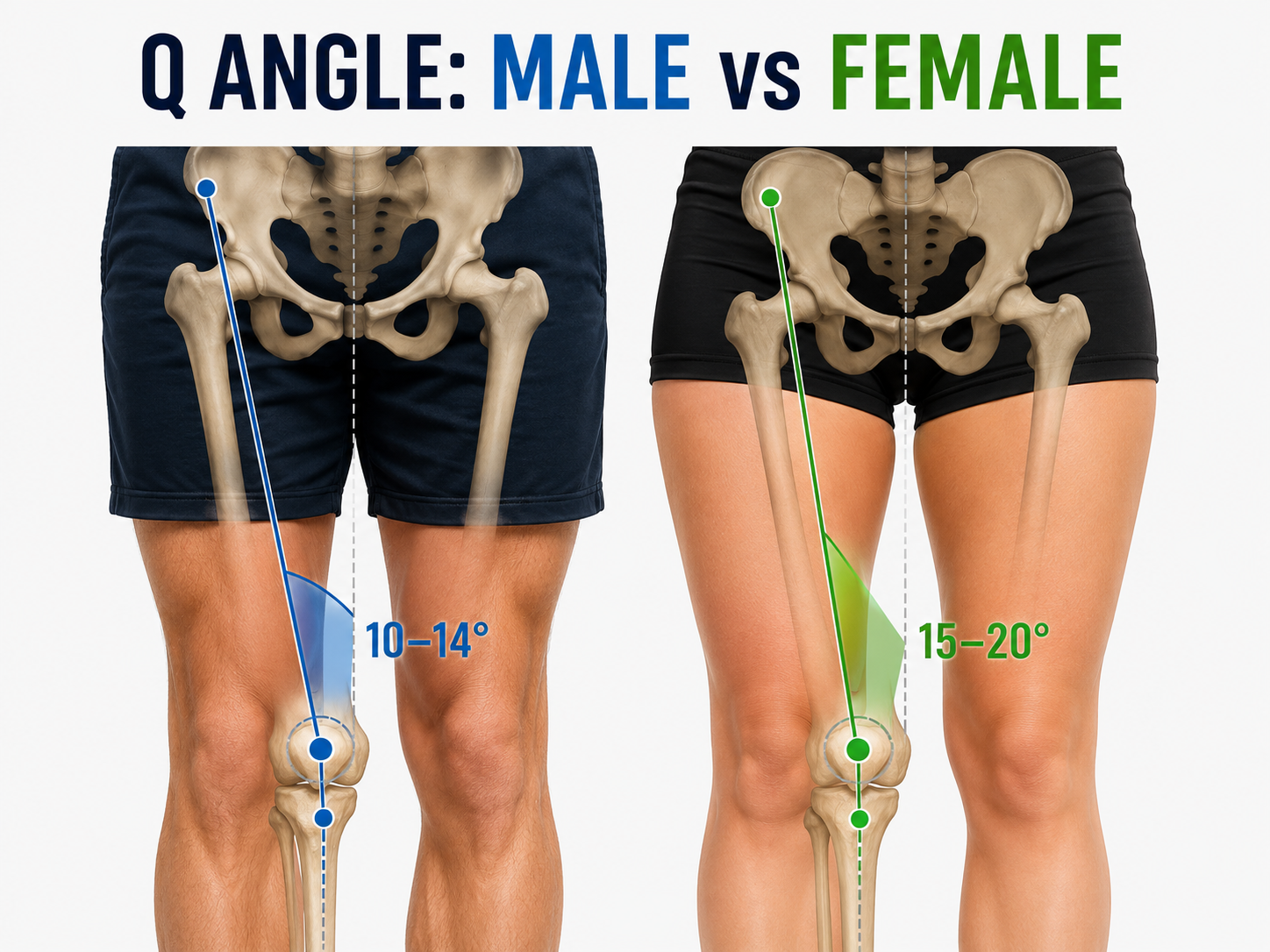
The Q-angle is the angle formed between the quadriceps muscle and the patellar tendon. Females have wider hips relative to their height, creating a larger Q-angle that pulls the kneecap — and the forces on the ACL — inward.
Female Q-angle averages 15–20°; male average is 10–14°. Every extra degree increases medial knee stress.

Knee valgus collapse during landing is the most common mechanism of ACL injury in female athletes.
The valgus problem
When a female athlete lands from a jump or cuts sharply, her knee frequently collapses inward — a movement pattern called dynamic valgus. This isn't weakness. It's a combination of anatomy, muscle activation patterns, and neuromuscular programming.
The inward collapse creates a perfect storm: the ACL is simultaneously stretched, rotated, and compressed. In a fraction of a second, forces that exceed the ligament's tensile strength cause a complete tear — often with no contact at all.
- • Knee caves inward
- • Hip drops on landing side
- • Foot pronates outward
- • ACL under maximum stress
- • Knee tracks over toes
- • Hip stays level
- • Foot neutral on landing
- • Forces distributed evenly
The estrogen effect
Estrogen receptors have been found in the ACL itself. Research shows that estrogen affects the mechanical properties of ligament tissue — and that these effects fluctuate throughout the menstrual cycle.
Studies have found that ACL injuries in female athletes cluster around the pre-ovulatory phase of the menstrual cycle, when estrogen levels peak. During this window, ligament laxity increases and neuromuscular control may be temporarily reduced.
Rising estrogen increases ligament laxity. Pre-ovulatory peak is the highest-risk window.
Estrogen surge at its peak. Studies show ACL injury rates are highest in the 2–3 days around ovulation.
Progesterone rises and partially counteracts estrogen's effect on laxity. Relative protective window.
Note: Research in this area is ongoing. Cycle-based training modifications are emerging but not yet standard practice. Consult a sports medicine professional for individualized guidance.
Growth spurt vulnerability
The peak risk window for ACL injury in female athletes is ages 14–17 — precisely when the adolescent growth spurt occurs. During this period:
- →Bones lengthen faster than muscles and tendons can adapt, reducing flexibility and neuromuscular coordination
- →The Q-angle widens as the pelvis broadens, increasing valgus stress on the knee
- →Estrogen levels rise sharply, affecting ligament laxity for the first time
- →Athletic training intensity often increases just as the body is most vulnerable
- →Neuromuscular patterns established before puberty may no longer match the new body geometry
Neuromuscular differences
Beyond anatomy and hormones, female athletes show measurable differences in how their nervous system activates muscles during athletic tasks:
Risk factors you can change
While anatomy and hormones can't be changed, the neuromuscular and biomechanical risk factors are highly trainable. Research shows prevention programs reduce ACL injury rates by up to 50%.
Strengthen the posterior chain
Targeted hamstring, glute, and hip abductor training corrects the quad-dominant pattern and gives the knee the braking force it needs.
See prevention exercisesRetrain landing mechanics
Plyometric and jump-landing drills teach the nervous system to keep the knee aligned over the toes — even under fatigue and game-speed pressure.
Soccer-specific drillsStart before the growth spurt
The most effective prevention window is ages 11–13, before the anatomical and hormonal changes of puberty amplify risk. Early training builds protective habits that persist.
Coach resources
Knowledge is the first step.
Now that you understand the why, explore the how — prevention exercises built specifically for female soccer athletes.